
Conventional spatial biology tells you where proteins are. Hawk Biosystems’ QF-Pro® technology tells you what they are doing. The first and only platform capable of spatially quantifying protein function in fixed tissue at nanoscopic resolution, QF-Pro® uses an antibody-based two-site assay combining amplified Förster Resonance Energy Transfer (FRET) and Fluorescence Lifetime Imaging Microscopy (FLIM) to measure functional proteomic events — protein-protein interactions, post-translational modifications, receptor heterodimerisation, protein-DNA interactions, and more — directly in FFPE tissue samples, at single-cell resolution, with quantitative precision that existing technologies cannot match. The signal is generated only when a functional event actually occurs, delivering readouts that are specific, reproducible, and directly biologically interpretable.
QF-Pro® operates as a biochemical ruler, making 1–10nm measurements within tissue samples to determine whether proteins are functionally engaged — not simply co-expressed or co-localised. This distinction is critical in oncology, immunology, and drug development, where the relationship between protein expression and protein function is frequently decoupled, and where treatment decisions and biomarker strategies built on expression data alone routinely fail. Published in the Journal of Clinical Oncology in 2023, QF-Pro® has demonstrated direct clinical value in immuno-oncology, accurately predicting patient response to immune checkpoint blockade in NSCLC where standard PD-L1 expression scoring did not. As Hawk Biosystems’ authorised Australian and New Zealand distributor, Decode Science can advise on applications, assay design, and getting started with the platform.
Quantify Protein Function. QF-Pro® delivers the layer of biological information that sits beneath expression data and directly drives cellular behaviour.
Proven Clinical Value in Immuno-Oncology. Stratifying patients by QF-Pro® could increase response rates by up to 280% and identify the approximately 47% of lung cancer patients.
High Specificity and Dynamic Range With a Higher Signal-to-Noise Ratio. Compatible with standard laboratory workflows and existing multiplex imaging infrastructure.
Validated off-the-shelf biomarkers include immune checkpoint interaction states, protein activation states (Akt, STAT3), and protein-protein interaction states (HER2/HER3, PKB/PDK1).
Identify your protein target of interest and application context. Validated off-the-shelf biomarkers are available for immune checkpoints, protein activation states, and protein-protein interaction states across multiple tumour types. Contact Decode Science for the current validated biomarker catalogue and to discuss custom target development if your assay of interest is not yet in the off-the-shelf repertoire.
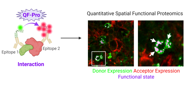
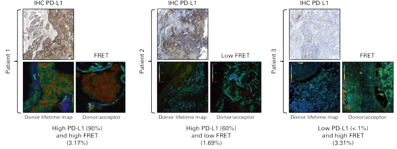
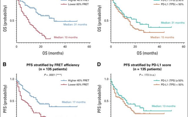
Image: Clinical PD-L1 images show the PD-L1 TPS expression, determined by IHC (SP263 Ventana Roche), on three patient samples. Below, the pseudocolored FLIM images are presented. The left panels show the lifetime maps of the FRET donor alone (no interaction). The right panels show the lifetime map of the FRET donor in the presence of the FRET acceptor. A reduction of donor lifetime in the presence of the acceptor (indicated by a change of pseudocolor from blue to green, yellow, and red depending on the FRET efficiency) represents the functional interaction of PD-1/PD-L1 within a patient sample. In these three examples, a discrepancy between FRET efficiency and PD-L1 expression is visible. DOI: 10.1200/JCO.22.01748
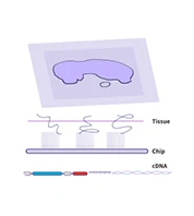
QF-Pro® is validated for FFPE tissue samples — compatible with archival and prospective tissue collections. Sample preparation follows standard immunofluorescence-based workflows, making QF-Pro® compatible with existing laboratory infrastructure without requiring additional specialist equipment.
Apply the two-site antibody labelling strategy to your tissue sections, using species-distinct primary antibodies against your two epitopes of interest, followed by QF-Pro® secondary labelling reagents. Assay protocols are available through Decode Science.


Image using Violet 3.0 — Hawk Biosystems’ dedicated FRET-FLIM imaging system. Violet 3.0 automatically measures donor-to-acceptor energy transfer, quantifying the functional event of interest and spatially mapping it across the tissue section. QF-Pro® software delivers reproducible, unbiased quantitative readouts without manual interpretation.
QF-Pro® outputs quantitative functional readouts with proven clinical cut-off values in immuno-oncology applications. Results can be interpreted in the context of existing validated clinical datasets, or used as the basis for new biomarker development and companion diagnostic programmes in collaboration with Hawk Biosystems.

ANZ Market Manager - Research Genomics
From protein presence to protein function — in FFPE tissue, at nanoscopic resolution.
QF-Pro® combines two established imaging modalities — FRET and FLIM — with a patented amplification step that makes their precision actionable in patient tissue for the first time. Two epitopes on the proteins of interest are labelled simultaneously with species-distinct antibodies. These are detected using QF-Pro® secondary labelling reagents — a green chromophore (donor) and an amplified red chromophore (acceptor). The Violet 3.0 imaging system automatically measures the distance between the donor and acceptor, based on the energy transferred between them. That distance is the functional readout. Signal is generated only when the functional event — the interaction, the modification, the activation state — is actually occurring.
Spatial proteomics has transformed how we understand tissue biology. But the information it delivers — protein localisation, co-expression, abundance — stops short of the questions that matter most in translational research and clinical decision-making. Whether a protein is present is not the same question as whether it is active, whether it is bound to its interaction partner, or whether a therapeutic antibody has engaged its target.
QF-Pro® is most relevant where that gap is widest:
PD-L1 expression alone predicts immunotherapy response poorly. QF-Pro® measures the actual PD-1/PD-L1 interaction state in tumour tissue, identifying responding patients that expression-based scoring misses — including the roughly 25% of NSCLC patients with low PD-L1 expression who nonetheless respond to checkpoint blockade.
Validated across ccRCC, NSCLC, melanoma, CRC, HNSCC, breast cancer, prostate cancer, and sarcoma. QF-Pro® biomarkers are designed to translate directly from research to companion diagnostic development, with a dedicated CDx partnership programme.
Directly measure whether a therapeutic antibody, ADC, or small molecule is engaging its target in tissue — not inferring it from downstream expression changes. Critical for mechanism-of-action studies and early efficacy signals.
Quantify intracellular PTMs including phosphorylation, methylation, glycosylation, and acetylation at single-cell resolution in tissue. Map pathway activation states spatially across heterogeneous tumour samples.


Multiplex spatial imaging for tissue sections; complements QF-Pro® for combined phenotypic and functional spatial profiling from the same FFPE tissue
Spatial transcriptomics at nanoscale resolution; pairs with QF-Pro® functional proteomics for multi-modal spatial characterisation of the tumour microenvironment
Ultrasensitive protein detection from serum and plasma; complements spatial functional proteomics with systemic biomarker measurement from liquid biopsy

Whole transcriptome single-cell RNA-seq from FFPE tissue; combines with QF-Pro® for transcriptomic and functional proteomic profiling from the same archival samples
Sánchez-Magraner, L., Gumuzio, J., et al. Journal of Clinical Oncology. 2023.

Miles J, Ward SG, Larijani B. PubMed.
QF-Pro® measures protein functional events in fixed tissue at nanoscopic resolution — including protein-protein interactions, post-translational modifications (phosphorylation, methylation, glycosylation, acetylation), receptor heterodimerisation, protein-DNA interactions, and single protein activation states. It does not measure protein expression or abundance alone — it measures whether proteins are functionally engaged.
Standard multiplex IF and IHC measure protein localisation and expression levels. QF-Pro® uses FRET-FLIM to measure the physical proximity of two labelled epitopes — generating signal only when the functional event is occurring. This means it reports on protein function, not protein presence, providing a fundamentally different and complementary layer of biological information.
QF-Pro® is validated for FFPE tissue samples, making it compatible with both prospective tissue collection and archival biobank material. Validated tissue types include NSCLC, ccRCC, melanoma, CRC, HNSCC, breast cancer, prostate cancer, sarcoma, and several normal tissue models.
Current validated off-the-shelf biomarkers include PD-1/PD-L1, CTLA-4/CD80, TIGIT/CD155, LAG-3/MHC II, TIM-3/Gal9 (immune checkpoint interaction states), Akt/PKB and STAT3 activation states, and HER2/HER3 and PKB/PDK1 interaction states. Contact Decode Science for the full current catalogue.
Yes. Hawk Biosystems works with clients to design and validate novel functional biomarkers for custom targets. Contact Decode Science to discuss custom assay development and the Hawk Biosystems partnership programme.
QF-Pro® data is acquired using Violet 3.0, Hawk Biosystems’ dedicated FRET-FLIM imaging system. Violet 3.0 automates the measurement and quantification process, making FRET imaging more accessible than conventional FLIM setups.
Yes. A peer-reviewed study published in the Journal of Clinical Oncology (2023) validated QF-Pro® for PD-1/PD-L1 interaction state measurement in 188 NSCLC patients, demonstrating that QF-Pro® readouts were highly predictive of response to immune checkpoint blockade therapy where standard PD-L1 TPS scoring was not.
We only need these information to serve you better. Decode Science respects your privacy and will never spam you with unrelated content.

Simply add your details below and get quick access to the brochure.
Simply add your details below and get quick access to the brochure.
Simply add your details below and get quick access to the brochure.